Arteries In Neck : The arteries in the chest, neck and brain are the most frequent arteries found to be abnormal in phace syndrome.
byAdmin•
0
Arteries In Neck : The arteries in the chest, neck and brain are the most frequent arteries found to be abnormal in phace syndrome.. There are two carotid arteries, one on the right and one on the left. Cervical bruits and hums may arise from neck arteries or veins, and may be innocuous findings or indicate underlying pathology. They are the carotid arteries, and they carry blood to the brain. What are the arteries of the chest, neck and brain? If one of them is narrowed or blocked, it can lead to a stroke.
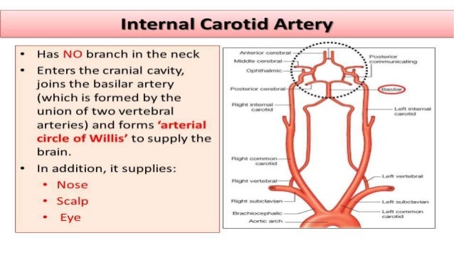
The vertebral arteries terminate by anastomosing together as the basilar artery. Oct 05, 2020 · in the neck, the carotid sheath (fibrous connective tissue) covers the common carotid artery, vagus nerve, and internal jugular vein. There are two carotid arteries, one on the right and one on the left. They are the carotid arteries, and they carry blood to the brain. These blood vessels can have abnormal shapes, sizes or paths through the neck and head.
Cervical Artery Dissection Health Information Bupa Uk from assets.bupa.co.uk The vein is the most lateral structure within the carotid sheath, followed by the nerve and then the artery, which is the most medial structure. The carotid arteries are major blood vessels in the neck that supply blood to the brain, neck, and face. Carotid artery stenosis is a narrowing in the large arteries located on each side of the neck that carry blood to the head, face and brain. The vertebral arteries terminate by anastomosing together as the basilar artery. There are two large arteries in the neck, one on each side. Over time, stenosis can advance to complete blockage of the artery. There are two carotid arteries, one on the right and one on the left. What are the arteries of the chest, neck and brain?
There are two carotid arteries, one on the right and one on the left.
Stomach, saclike expansion of the digestive system, between the esophagus and the small intestine; The carotid arteries are major blood vessels in the neck that supply blood to the brain, neck, and face. The arteries in the chest, neck and brain are the most frequent arteries found to be abnormal in phace syndrome. These blood vessels can have abnormal shapes, sizes or paths through the neck and head. Blood is carried to the brain through blood vessels called arteries. Carotid artery stenosis is a narrowing in the large arteries located on each side of the neck that carry blood to the head, face and brain. The stomach serves as a temporary receptacle for the storage and mechanical distribution of food before it is passed into the intestine. Oct 05, 2020 · in the neck, the carotid sheath (fibrous connective tissue) covers the common carotid artery, vagus nerve, and internal jugular vein. There are two large arteries in the neck, one on each side. Through their course, they give off several meningeal, muscular and spinal branches for the nearby structures. What are the arteries of the chest, neck and brain? The vertebral arteries terminate by anastomosing together as the basilar artery. Blood is pumped from the ventricles into large elastic arteries that branch repeatedly into smaller and smaller arteries until the branching results in microscopic arteries called arterioles.
There are two carotid arteries, one on the right and one on the left. May 31, 2021 · the vertebral arteries ascend through the neck inside the transverse foramina of the cervical vertebrae, all the way to the brain. Cervical bruits and hums may arise from neck arteries or veins, and may be innocuous findings or indicate underlying pathology. Through their course, they give off several meningeal, muscular and spinal branches for the nearby structures. Blood is carried to the brain through blood vessels called arteries.
F38loiyngfpqcm from image.slidesharecdn.com Blood is pumped from the ventricles into large elastic arteries that branch repeatedly into smaller and smaller arteries until the branching results in microscopic arteries called arterioles. Carotid artery stenosis is a narrowing in the large arteries located on each side of the neck that carry blood to the head, face and brain. Doctors can test for a narrowed carotid artery, but it's usually not a good idea. Through their course, they give off several meningeal, muscular and spinal branches for the nearby structures. Over time, stenosis can advance to complete blockage of the artery. Oct 05, 2020 · in the neck, the carotid sheath (fibrous connective tissue) covers the common carotid artery, vagus nerve, and internal jugular vein. In fact, the test may do more harm than good. There are two carotid arteries, one on the right and one on the left.
What are the arteries of the chest, neck and brain?
Cervical bruits and hums may arise from neck arteries or veins, and may be innocuous findings or indicate underlying pathology. They are the carotid arteries, and they carry blood to the brain. The vertebral arteries terminate by anastomosing together as the basilar artery. In fact, the test may do more harm than good. Carotid artery stenosis is a narrowing in the large arteries located on each side of the neck that carry blood to the head, face and brain. If one of them is narrowed or blocked, it can lead to a stroke. May 31, 2021 · the vertebral arteries ascend through the neck inside the transverse foramina of the cervical vertebrae, all the way to the brain. What are the arteries of the chest, neck and brain? Oct 05, 2020 · in the neck, the carotid sheath (fibrous connective tissue) covers the common carotid artery, vagus nerve, and internal jugular vein. Stomach, saclike expansion of the digestive system, between the esophagus and the small intestine; These blood vessels can have abnormal shapes, sizes or paths through the neck and head. The vein is the most lateral structure within the carotid sheath, followed by the nerve and then the artery, which is the most medial structure. Systemic arteries transport oxygenated blood from the left ventricle to the body tissues.
The stomach serves as a temporary receptacle for the storage and mechanical distribution of food before it is passed into the intestine. There are two large arteries in the neck, one on each side. In fact, the test may do more harm than good. What are the arteries of the chest, neck and brain? Over time, stenosis can advance to complete blockage of the artery.
Carotid Artery Disease Stenting Vs Endarterectomy British Journal Of Anaesthesia from els-jbs-prod-cdn.jbs.elsevierhealth.com Through their course, they give off several meningeal, muscular and spinal branches for the nearby structures. Carotid artery stenosis is a narrowing in the large arteries located on each side of the neck that carry blood to the head, face and brain. In fact, the test may do more harm than good. Blood is pumped from the ventricles into large elastic arteries that branch repeatedly into smaller and smaller arteries until the branching results in microscopic arteries called arterioles. The vertebral arteries terminate by anastomosing together as the basilar artery. Neck auscultation is commonly indicated for initial evaluation of stenotic or embolic cerebrovascular symptoms, or as part of a comprehensive physical examination in asymptomatic patients at risk for atherosclerosis. The carotid arteries are major blood vessels in the neck that supply blood to the brain, neck, and face. Cervical bruits and hums may arise from neck arteries or veins, and may be innocuous findings or indicate underlying pathology.
The arteries in the chest, neck and brain are the most frequent arteries found to be abnormal in phace syndrome.
Doctors can test for a narrowed carotid artery, but it's usually not a good idea. Stomach, saclike expansion of the digestive system, between the esophagus and the small intestine; The stomach serves as a temporary receptacle for the storage and mechanical distribution of food before it is passed into the intestine. Oct 05, 2020 · in the neck, the carotid sheath (fibrous connective tissue) covers the common carotid artery, vagus nerve, and internal jugular vein. The vein is the most lateral structure within the carotid sheath, followed by the nerve and then the artery, which is the most medial structure. Systemic arteries transport oxygenated blood from the left ventricle to the body tissues. If one of them is narrowed or blocked, it can lead to a stroke. Blood is carried to the brain through blood vessels called arteries. Neck auscultation is commonly indicated for initial evaluation of stenotic or embolic cerebrovascular symptoms, or as part of a comprehensive physical examination in asymptomatic patients at risk for atherosclerosis. These blood vessels can have abnormal shapes, sizes or paths through the neck and head. They are the carotid arteries, and they carry blood to the brain. Through their course, they give off several meningeal, muscular and spinal branches for the nearby structures. May 31, 2021 · the vertebral arteries ascend through the neck inside the transverse foramina of the cervical vertebrae, all the way to the brain.